Key Takeaways
- Keratoconus is a condition where the cornea (the clear front surface of the eye) thins and bulges outward into a cone-like shape.
- This irregular shape causes blurred or distorted vision that glasses may not fully correct.
- Symptoms often begin in the teenage years or early adulthood and may worsen over time.
- Diagnosis involves a detailed eye examination, including corneal mapping.
- Management options range from special contact lenses to surgical procedures depending on severity.
Most people think of blurry vision as something that can be fixed with a new pair of glasses. But what if glasses don’t solve the problem? For some, the cause is keratoconus — a progressive condition where the normally round cornea becomes thinner and bulges outward into a cone shape.
This change in shape bends light unevenly, making vision blurry, distorted, or sensitive to glare, often affecting both eyes to varying degrees.
What Is Keratoconus?
Keratoconus is an eye condition that gradually alters the cornea’s shape. Instead of remaining round, the cornea weakens and starts to thin, forming a cone-like bulge. This makes it harder for the eye to properly focus light onto the retina, leading to vision problems.
Causes and Risk Factors
The exact cause is not fully understood, but several factors are linked to keratoconus:
- Genetics – It can run in families.
- Eye rubbing – Frequent, vigorous eye rubbing has been associated with progression.
- Underlying conditions – Such as asthma, allergies, or connective tissue disorders.
Symptoms to Watch Out For
Common symptoms include:
- Blurry or distorted vision
- Increased sensitivity to light and glare
- Frequent changes in glasses prescription
- Double vision in one eye (monocular diplopia)
- Difficulty seeing clearly at night
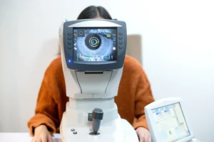
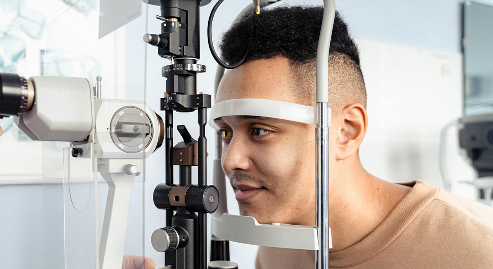
Diagnosis
An eye specialist may use several tests to confirm keratoconus, including:
- Corneal topography – a detailed map of the cornea’s shape and thickness.
- Slit-lamp examination – to look for thinning or scarring.
- Keratometry – measures the curvature of the cornea.
Treatment and Management
Treatment depends on the severity of the condition:
- Glasses or soft contact lenses – may help in the early stages.
- Specialty contact lenses – such as rigid gas permeable, scleral, or hybrid lenses to improve vision when regular lenses no longer work well.
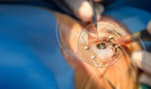
- Corneal cross-linking – a procedure that strengthens the cornea to slow or halt progression.
- Corneal transplant – considered in advanced cases where other treatments are not sufficient.
Living With Keratoconus
With proper monitoring and treatment, many people with keratoconus can maintain functional vision. Regular check-ups are important to track changes in the cornea and adjust treatment as needed.
When to See an Eye Specialist
If you notice that your vision remains blurred despite updated glasses or lenses, or if lights at night seem unusually distorted, it’s best to consult an eye specialist. Early detection of keratoconus can open up more options for managing the condition effectively.
Key Takeaways Recap
- Keratoconus occurs when the cornea thins and bulges outward.
- It leads to blurry, distorted vision that glasses may not fully correct.
- Symptoms include glare, frequent prescription changes, and poor night vision.
- Diagnosis relies on corneal mapping and eye examination.
- Management may involve specialty lenses, corneal cross-linking, or surgery in advanced cases.
If you’re experiencing ongoing blurry vision or suspect keratoconus, consider booking an eye examination at London Eye & Retina for a detailed assessment.