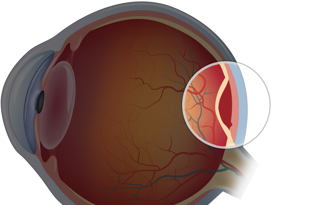
A Vision Emergency That Requires Prompt Attention
Retinal detachment occurs when the retina separates from the back of the eye, disrupting the visual signals it sends to the brain. It can develop suddenly and may cause warning signs such as flashes of light, a sudden increase in floaters, blurred vision, or a dark shadow or curtain spreading across part of your field of vision.
Because retinal detachment can cause permanent vision loss if treatment is delayed, these symptoms should be assessed urgently by an eye specialist. A detailed retinal examination can identify the extent of the detachment and determine the most appropriate retina macula treatment to help protect your vision.
At London Eye & Retina, we treat retinal detachment as a sight-threatening emergency. Early diagnosis and timely surgery can make all the difference in preserving your vision.
What Is Retinal Detachment?
The retina is the thin layer of light-sensitive tissue lining the back of the eye. When it separates from the underlying layer that provides oxygen and nutrients, the retina can no longer function properly, resulting in vision loss in the affected area.
There are three main types:
- Rhegmatogenous (most common): Caused by a tear or hole in the retina, allowing fluid to seep underneath
- Tractional: Often seen in diabetic eye disease, where scar tissue pulls the retina away.
- Exudative: Caused by fluid buildup due to inflammation, injury, or tumours without a tear
What Causes It Retinal Detachment?
- Age-related vitreous changes (common in people over 50)
- High myopia (severe short-sightedness)
- Trauma to the eye
- Previous eye surgery (e.g., cataract surgery)
- Family history of retinal detachment
- Diabetic retinopathy (tractional type)
Symptoms to Watch For Retinal Detachment
Retinal detachment is typically painless but can cause dramatic visual symptoms, such as:
- Sudden onset of floaters (black spots or threads)
- Flashes of light in one eye
- A shadow or “curtain” over part of the vision
- Sudden drop in visual sharpness or darkening of vision
These symptoms can come on quickly and worsen over hours or days.
How Is Retinal Detachment Diagnosed?
At London Eye & Retina, we perform a detailed eye examination to assess the retina using:
- Dilated fundus examination
- Optical Coherence Tomography (OCT) to evaluate retinal structure
- Ultrasound (B-scan) is used in cases where the retina is not easily visible due to bleeding or cloudiness
Early detection allows for better planning of the surgical repair.
Treatment Options For Retinal Detachment
Surgery is required to reattach the retina and prevent further vision loss. The type of surgery depends on the type, location, and extent of the detachment:
- Vitrectomy: Removes the vitreous gel and uses a gas bubble or silicone oil to flatten the retina
- Scleral Buckle: A silicone band is placed around the eye to relieve traction and support the retina
- Pneumatic Retinopexy: A gas bubble is injected into the eye to push the retina back into place, combined with laser or cryotherapy
Your surgeon will recommend the most suitable method based on your eye’s condition.
Recovery and Long-Term Care
Post-surgery, patients may need to maintain a specific head position to help the retina heal. Vision recovery can take several weeks to months and may not always return to pre-detachment levels, especially if the macula was involved.
Follow-up care includes regular monitoring, managing any pressure changes, and watching for complications such as re-detachment or cataract formation.
Act Fast to Save Your Sight
Retinal detachment is a medical emergency. The sooner it’s treated, the better the chance of preserving vision. Don’t wait; prompt action can help prevent permanent damage.
Schedule a consultation with Dr. James Ng at London Eye & Retina and explore your options for clearer, sharper sight.
Let us help you respond quickly and restore your retinal health.